Center News & Funding, Research, Prevention & Control, Clinical, Technology
Sep 27, 2024
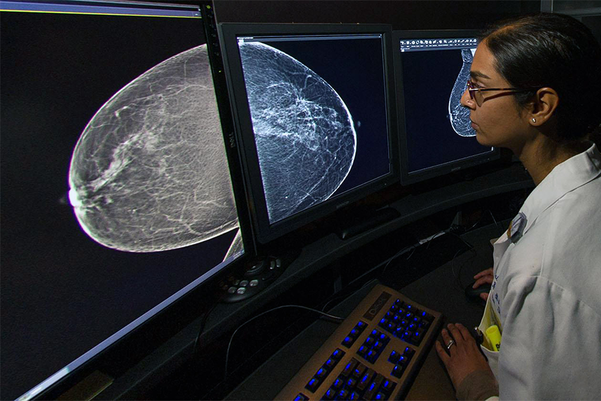 Priti Shah, M.D., director of Breast Imaging at VCU Health and VCU Massey Comprehensive Cancer Center, shares insight into new FDA mammogram rules.
About half of women who are 40 or older in the United States have dense breast tissue. This type of tissue not only puts them at higher risk of developing breast cancer – it also makes it harder for clinicians to detect it in mammograms.
In September, the U.S. Food and Drug Administration released new rules that require radiologists to directly send patients a summary after having a mammogram, notifying them of their breast tissue composition and other findings.
“Previously, patients were only informed about their breast density if their state laws required it and if they had dense breast tissue. This information about breast density has always been sent to clinicians in a more formal report,” said Priti Shah, M.D., director of Breast Imaging at VCU Health and VCU Massey Comprehensive Cancer Center.
The report will not be formal or highly clinical, like the one sent to providers, Shah says. Instead, it will be written in more common language so it’s easier for patients and their loved ones to read.
“The change provides uniformity and standardization for patients and can empower them to make informed decisions about cancer screenings and prevention,” she added.
Shah recently spoke with VCU Health News about the latest changes in mammography reports and the importance of early detection to treat cancer.
A mammogram is a low dose x-ray of the breast. Mammograms are currently the gold standard for early detection of breast cancer.
There is an overwhelming amount of scientific evidence that when women over age 40 get mammograms annually, deaths from cancer decrease by up to 40%. This is in large part because mammograms are excellent at showing early-stage breast cancer (Stage 0) or ductal carcinoma in situ (DCIS) — when cancer is still confined to the milk duct and hasn't invaded surrounding breast tissue to form a "lump," or spread to lymph nodes or other organs.
When cancer is detected at its earliest stages, the patient is more likely to have a better prognosis and chances for survival. Also, our care teams at Massey may be able to pursue less aggressive cancer treatments.
Breast density is a ratio that refers to the amount of fibrous and glandular – or "fibroglandular” – tissue in the breast compared to fatty tissue. This ratio varies person-to-person based on genetics and hormones.
The fibroglandular tissue is brighter white on a mammogram, making it harder to see through, while the fatty tissue is gray and more transparent. Lumps, both cancerous and non-cancerous (benign), tend to be the same shade of white as normal fibroglandular tissue. So, the denser the breast tissue, the harder it is to find small cancers as they are hidden or masked by the normal tissue.
Higher breast density may also increase one's risk for breast cancer. A patient is said to have dense tissue if less than 50% of their tissue is fibroglandular on a mammogram.
There are two mammography reports. One is the "formal" report that is sent to the clinician who requested the mammogram. This will have detailed information about the patient's breast density, what prior studies it was compared to, any important findings, such as suspicious lumps or calcium deposits, or other signs of breast cancer, and a recommendation for a plan. The recommendation or plan may be as straightforward as returning for a screening in one year if all is normal or benign, or to return for further imaging.
This report will also include a numerical score, called the BI-RADS (Breast Imaging Reporting and Data System) Assessment, which classifies the results into categories ranging from normal (1) to highly suspicious (5). This, and other parts of the BI-RADS, provides radiologists a standardized way of communicating mammogram results by using the same terminology, to make their overall interpretation of the mammogram, and any follow up recommendations, clear.
The other report is a letter sent directly to patients in plain language. This is unique to breast imaging; it's the only radiology test for which a report is required to be sent directly to the patient even before the days of patient portals. This letter provides the patient with information about whether their breast tissue is dense or not dense, as well as an overview of the results as interpreted by the radiologist, and a recommendation for follow up.
The FDA now requires that all patients are told in their results letter whether they have dense tissue or not and what that means. Additionally, they will be advised to talk with their doctor about additional screening tools in light of their other risks for breast cancer. Radiologists must also use specific wording about this as directed by the FDA to ensure consistency in what patients are being told.
Here are some examples of the language that may be used in reports about breast density:
Mammograms are still the gold standard for Stage 0 cancers in all breast densities. Dense breast tissue can hide small invasive cancers that can spread if undetected and increase one's risk for breast cancer. Given the limitations of mammography in dense tissue, additional screening tools, used in conjunction with mammograms, can help increase the chances of finding a small cancer when it is most treatable.
We provide breast MRI and/or ultrasound in conjunction with mammography for additional screening, as ordered by a referring provider. There is a lot of variation with regard to insurance coverage, so we encourage patients to call their insurance provider and inquire about the specifics of their policy and what out-of-pocket costs they may incur.
VCU Breast Imaging staff see patients with both cancer and non-cancerous breast conditions. We use every in-person encounter to discuss breast density with the patient, addressing many of the same questions and concerns explained above.
Outside the walls of Massey, we also participate in patient education in the community, such as health fairs and wellness events. To see where we'll be next, explore our calendar of events.
Written by: Sara McCloskey
Clinical, Research, Center News & Funding
Get C-SMART: Massey neuropsychologist seeks to enhance brain wellness in neuro-oncologySep 23, 2024
Center News & Funding, Community Engagement & Health Equity, Massey 50
Massey hosts congresswoman, highlights efforts to reduce cancer burden for all VirginiansSep 16, 2024
Research
Undergraduate student embraces the chance to write her own story in cancer researchAug 30, 2024

Treatments in clinical trials may be more effective or have fewer side effects than the treatments that are currently available. With more than 200 studies for multiple types of cancers and cancer prevention, Massey supports a wide array of clinical trials.

Massey supports hundreds of top cancer specialists serving the needs of our patients. Massey’s medical team provides a wealth of expertise in cancer diagnosis, treatment, prevention and symptom management.